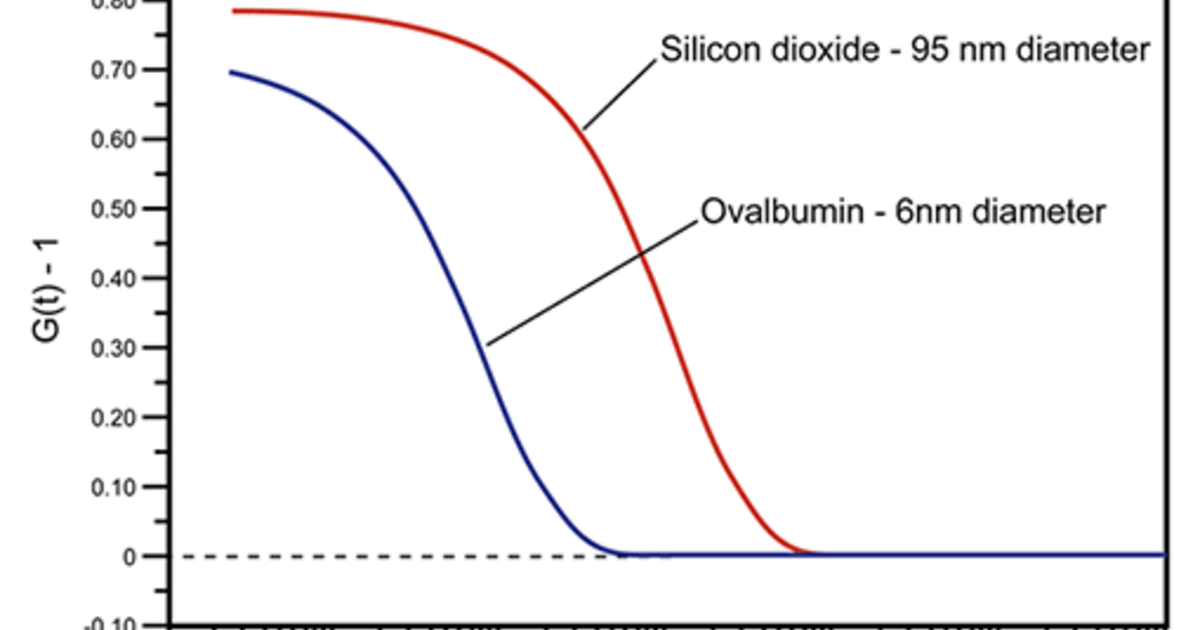
The correlation function g(t) has been systematically measured as a function of the fibrinogen concentration in solutions at a fixed pH 8, activated by 400 nM of thrombin. Fibrinogen is a protein present in vertebrate body fluids, which is capable of forming gels because of an initial activation and a successive polymerization process. Quasi-elastic light scattering measurements of the normalized time-dependent density correlation function, g(t), have been carried out on fibrin networks formed at T equals 25 degree(s)C from fibrinogen solutions. From these fits, we conclude (1) the stretched exponential fit is worst if η μ < η, (2) models predicting a stretched exponential offer no explanation for η μ < η, and (3) the experimentally determined values for v compare well to other experimental data but not to theoretical predictions. The sphere diffusion constant data was fit to a stretched exponential of the form D/D o equals exp(-αc v) which is predicted by several models for the diffusion of spheres in polymer solutions. Microviscosities (η μ ) as low as 1/2 of the solution viscosity (η) were found. The sphere diffusion constants in the composite liquids were measured as a function of the rod concentration by dynamic light scattering. The sizes of the macromolecular components in each solution were chosen such that the sphere radius/rod length ratio decreased from CL1 to CL5. The present results suggest a simple model for organization of condensed dsDNA chromosomes in icosahedral viruses.įive composite liquid solutions (CL1-5) were prepared consisting of silica spheres and the rigid rod polymer poly(γ-benzyl α,L-glutamate) (PBLG) in the polar organic solvent dimethylformamide. We find no evidence in the Raman spectrum of specific intermolecular interactions involving capsid protein and major groove sites of the packaged DNA. The findings are compared with structural changes in protein subunits of the P22 viral capsid, attendant with capsid expansion and DNA packaging. Here we describe the use of digital difference Raman methods to probe structures of the double-stranded (ds) DNA genome of the icosahedral virus P22 in packaged and unpackaged states. Raman spectroscopy is particularly well suited to investigating assembly mechanisms of viruses and conformations of their nucleic acid and protein constituents. The Raman technique is a valuable complement to high-resolution structure methods applied to biological molecules in the crystal (x-ray crystallography) and to static and dynamic light scattering methods applicable to solutions. Laser Raman scattering is the method of choice for probing macromolecular structures and interactions in complex biological assemblies. Control experiments with latex particles in mucin and vesicles in other proteins showed no change in size, implying that the fusion of vesicles is due to vesicle-mucin interactions.

Repeated measurements of the intensity correlation functions after adding mucin to a suspension of vesicles showed a two-fold increase in the hydrodynamic radius of the vesicles over a period of three hours after which the vesicle size stayed constant. Similar methods were used to investigate the interaction of gall bladder mucin with cholesterol-phospholipid vesicles. The results showed that at low pH (below 4) solutions of gastric mucin contain very large supra-molecular aggregates, with diffusion constants 100 times slower than those of the 2 X 10 6 molecular weight glycoprotein of mucin. Intensity autocorrelation functions measured on purified mucin solutions under varying experimental conditions were analyzed by Laplace inversion methods.

Dynamic light scattering was applied to study aggregation phenomena in mucin, the glycoprotein responsible for the visco-elastic properties of mucus which is found as a lining on most epithelial cell surfaces.


 0 kommentar(er)
